
With the rapid technological advancement, science has evolved manifolds over the recent decades. With new discoveries, more is being uncovered every second. Moreover, the horizon of technology has spread its wings into almost every field. Grunstein and Hogness are credited for creating the first DNA array using the method of colony hybridization. The concept of microarrays has also caught a lot of attention lately and with its development, it is now being used in a number of different ways including the treatment of cancer.
The microarray technology makes observation of a multitude of genes at the same time convenient. It makes use of the knowledge that the sequence of a complimentary nucleotide of a DNA matches with its counterpart and will come together.
Microarray is primarily a grid-type disk or surface on which miniscule patterns of the genetic material are laid out. One spot represents a certain type of gene. The types of genes in each slot are backed up on a computer to keep track of which gene is placed where. How it becomes useful for medicine is that this plate can determine which spot or gene is responsible or becomes active during a certain medicine is taken.
You can take the example of a bacterium to understand the concept. So, for instance, you want to know which gene predominantly reacts to an anti-biotic, you get the microarray of that particular bacterium and break it open. Then you take out the RNA components from two different media, one plain and the other with a low anti-biotic level. Here the level of antibiotic is just enough to activate resistance of the bacterium and simultaneously ensure that the dose is so dilute that it does not kill the bacterium.
The next step involves tagging a small sample from each media. The RNA of the plain one could be dyed green and the anti-bacteria one could be dyed red. Then, this RNA is poured onto the microarray to combine to their complimenting strands and left for a while so that they can find their companions. Thus, during this time, the RNA moves around, bumping into the different types of DNAs present on the chip until it finds THE one. RNAs that fail to stick to DNAs are then washed off.
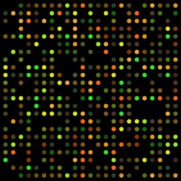
Then, a laser scanner is used to spot the dyes. An image of the pattern is taken which is used for analysis. There will be three different colours detected: green, yellow and red. The green dyes would point towards genes that do not have antibiotic resistance. The yellow ones would be RNAs from both untreated and anti-biotic treated samples that attached themselves to their corresponding DNAs. Thus these represent genes that are active irrespective of the presence or absence of antibiotics. The red ones indicate genes that are primarily active only in bacteria treated with anti-biotic.
The amount of brightness of each spot is directly proportional to their level of activity. These red genes would then be of interest to the scientist so they can find a way to weaken them or create a stronger antibiotic that acts upon them as well.
With this technology, research can now be done on a quicker pace.
Samantha Stinson is a microbiologist and encourages the use of microarray consumables. She has had an amazing relation concerning the equipment with http://www.arrayjet.co.uk/.